
Thematic research data commons is:
People
Reducing Deaths from Air Pollution
Exploreabout Reducing Deaths from Air Pollution
Three million people around the world – more than 33,000 of whom are in Australia – are living with multiple sclerosis (MS). It is a disease that damages nerves and disrupts communication between the brain and the body. This can result in difficulty walking, vision changes and various other ailments.
There is currently no cure for MS, but neurologists around the world are collaborating to better understand it and improve healthcare for patients.
The MSBase Registry is a global online registry for MS researchers, dedicated to sharing, tracking and evaluating data on outcomes in MS and other neuro-immunological diseases. Since its launch in 2004, it has become the largest organised repository of longitudinal MS patient data, with over 85,000 patient records from health facilities in 45 countries.
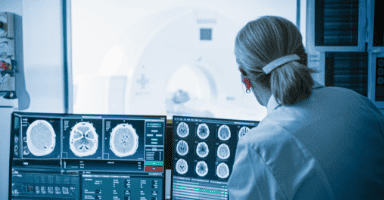
Now a new global platform – the MSBase Imaging Repository (MSBIR) – is further facilitating international collaboration in MS research by enabling secure sharing and analysis of de-identified clinical images of MS, such as MRI scans. Integrated with the MSBase Registry, it was developed in a partnership between The University of Sydney’s Brain and Mind Centre, MSBase and the Sydney Neuroimaging Analysis Centre (SNAC), and one of its building blocks is the ARDC-supported Australian Imaging Service (AIS).
The AIS is a national platform for secure management, analysis and informatics of mainly biomedical and veterinary imaging data. It is led by The University of Sydney in partnership with and with co-investment from the ARDC, the National Imaging Facility and 10 other universities and research organisations.
“Our mission is to increase research reproducibility and drive the adoption of innovative but trusted analysis techniques. We aim to create a unified service underpinning all imaging research conducted by Australians, both nationally and abroad, on which more specific research and development programs can be built,” said Dr Ryan Sullivan, who leads the AIS.
The AIS operates as a federation. It co-maintains a central set of software repositories with each partner institution operating their own node according to their local governance, infrastructure and cost structures.
The AIS integrates with imaging devices in a hub-and-spoke model. Each node integrates their local academic and clinical equipment, and data can be transferred between nodes to facilitate multi-site studies.
Our mission is to increase research reproducibility and drive the adoption of innovative but trusted analysis techniques. We aim to create a unified service underpinning all imaging research conducted by Australians, both nationally and abroad, on which more specific research and development programs can be built.
Dr Ryan Sullivan, Head, Australian Imaging Service
By adopting and standardising user authentication, instrument integration, data ontologies and mature software tools, the AIS lets researchers and facilities spend more time on innovation. It also allows reuse of national datasets by building a provenance trail from image capture to manipulation.
Taking a data-centric computing approach, the AIS integrates analysis and informatics directly into data management. This is broken into 4 key capability areas, the first 2 of which are live now:
The AIS team is keenly aware of the need for international collaboration in imaging research. This is precisely the case with research into MS, a global challenge requiring global solutions like the MSBIR.
For the MSBIR project, the AIS collaborated with Radiologics, which designed a federated system that would allow patient data to be domiciled in its home country while coordinating access for specialists from a single point that dynamically aggregates data for analysis.
Thanks to the cloud-native design of the AIS, the Australian, US, EU and lead MSBIR AIS nodes are in full production. Each node is accompanied by the Sydney Neuroimaging Analysis Centre’s TORANA software, which handles seamless data ingestion and custom integration with the MSBIR analysis engine to provide AI-based, quantitative imaging metrics. By providing secure, scalable and audited infrastructure across multiple jurisdictions around the world for long-term clinical image management, the AIS enables the large-scale analytics needed by global MS researchers for new insights into the disease.
“Going forward, our partnership with the AIS will further optimise the successful workflow and ongoing global implementation of the MSBIR,” said Professor Michael Barnett and Dr Heidi Beadnall, leaders of the MSBIR Project Control Board. It is also opening the way for global, more widely accessible AIS nodes to facilitate partnerships between Australia and the rest of the world.
The conclusion of the ARDC AIS project is the start of the AIS platform. We currently support 398 users across 279 projects and have a number of large programs that we are partnering with to support, such as the ACRF Australian Centre of Excellence in Melanoma & Diagnosis and recently announced Medical Research Future Fund projects.
Dr Ryan Sullivan, Head, Australian Imaging Service
The AIS team is now working with other ARDC-supported projects, such as the Australian Electrophysiology Data Analytics Platform, the Veterinary and Animal Research Data Commons, and the Australian Characterisation Commons at Scale.
“The conclusion of the ARDC AIS project is the start of the AIS platform. We currently support 398 users across 279 projects and have a number of large programs that we are partnering with to support, such as the ACRF Australian Centre of Excellence in Melanoma & Diagnosis and recently announced Medical Research Future Fund projects,” said Dr Sullivan.
Learn more about the AIS.
The AIS (doi.org/10.47486/PL102) was a partnership of the ARDC, the National Imaging Facility, The University of Sydney, Macquarie University, Queensland Cyber Infrastructure Foundation, Queensland University of Technology, The University of Queensland, UNSW Sydney, Neuroscience Research Australia, The University of Western Australia, and SAHMRI. Swinburne University, Monash University and The Florey advised on the project.
The ARDC is continuing to address the health and medical data challenges associated with secure data access, data integration and advanced analysis through the People Research Data Commons.
Written by Jason Yuen (ARDC). Edited by Mary O’Callaghan. Reviewed by Jo Savill (ARDC), Kerry Levett (ARDC), Adrian Burton (ARDC), Keith Russell (ARDC), Dr Heidi Beadnall (MSBIR), Professor Michael Barnett (MSBIR) and Dr Ryan Sullivan (AIS).